|
||||||||||||||||
Diffusion of proteins through thin hydrogel films and through cell glycocalyx
- Concept: hydrogel structure creates steric hindrance to the diffusion of proteins. In addition, some hydrogel polymer chains are "sticky" to the protein and the weak association slows protein diffusion even further. Protein diffusion in thin hydrogel films can be measured directly using fluorescence correlation spectroscopy.
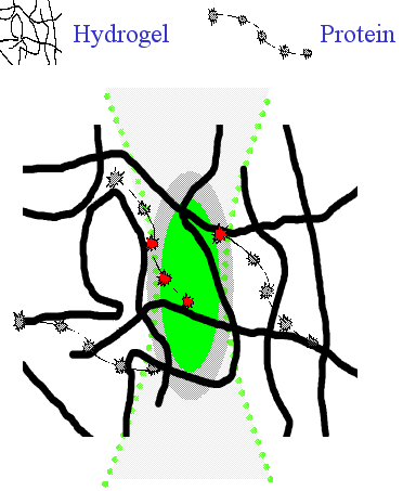
Schematics of the hydrogel structure with diffusing protein. Superimposed to the structure is the observation volume of FCS (not drawn to scale).
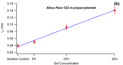
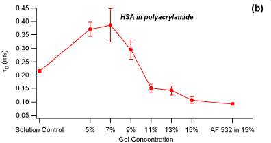
- Characteristic diffusion times for a small dye and albumin scale differently with polyacrylamide gel concentration. Polyacrylamide gel is non-sticky for protein, but at high gel concentrations protein can not get into the gel.
Characteristic diffusion times obtained by fitting experimental FCS data to a 3D model for a small dye (AlexaFluor 532) and human serum albumin (labeled with AF532) shown as a function of gel concentration. The lower values of characteristic diffusion times indicate larger diffusivity. (The drop of characteristic diffusion times for HSA in higher polyacrylamide concentration turned out to be due to a small amount of free dye in protein solution!). (from A. Stevens, MS Thesis, U of Utah).
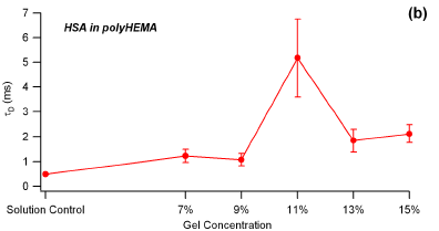
- Same protein in a "sticky' hydrogel, poly(HEMA), show different diffusivities.
Characteristic diffusion times obtained by fitting experimental FCS data to a 3D model for human serum albumin (labeled with AF532) shown as a function of polyHEMA concentration. (from A. Stevens, MS Thesis, U of Utah).
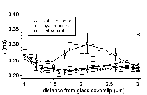
- Same FCS methodology can be aplied to study protein diffusion through cell glycocalyx. In the example below, the characteristic diffusion times for HSA-AF532 conjugate have been measured by scanning the FCS observation volume through the endothelial cell before and after the glycocalyx has been treated with hyaluronidase enzyme that degraded part of the glycocalyx structure.
Characteristic diffusion times for albumin (labeled with AF532) within and above the bovine lung microvascular endothelial cells. At the distances of approx. 2 µm from the cell-substrate interface one observes the slowing down of protein diffusion due to the interactions with glycocalyx. Once the hyaluronidase removes hyaluronan component of the glycocalyx, albumin diffusion profile returns to solution control values (from Stevens AM, Hlady V, Dull R , Fluorescence Correlation Spectroscopy Can Probe Albumin Dynamics Inside Lung Endothelial Glycocalyx. Am J Physiol Lung Cell Mol Physiol 2007 May 4;(ePrint)).
.